
Research
Skin as a Sensor for Disease
Skin is visible, accessible, and easy to study over time. We use genetic, molecular, imaging, and digital tools to find disease signals in the skin and measure how they change.
These approaches guide our work in skin cancer, neurofibromatosis, autoimmune disease, and other conditions where the skin reveals important information about health.
Genetic Signals
Skin as a Genetic Sensor
Genetic changes shape a person's risk of skin disease and how tumors behave. We study patients, tumors, and skin findings to identify inherited and acquired variants linked to skin cancer, neurofibromatosis type 1, and treatment response.
Selected projects
- Identifying genetic factors associated with skin cancer risk
- Studying genes that influence tumor development and treatment response
- Defining genetic factors associated with neurofibroma burden in NF1
Molecular Signals
Skin as a Molecular Sensor
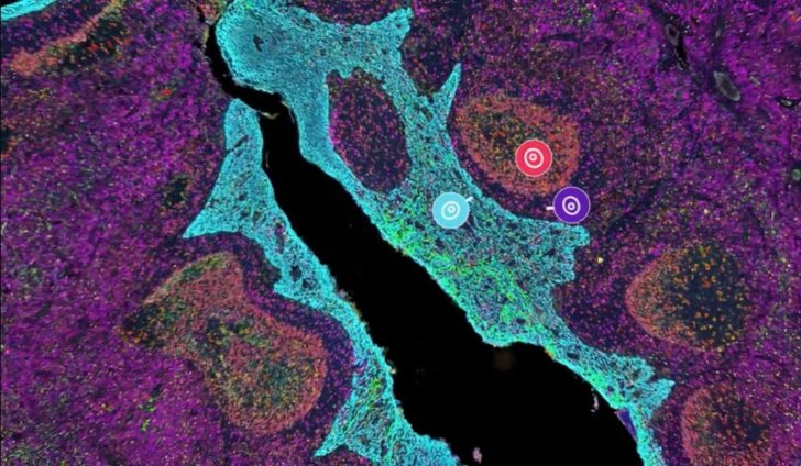
Skin contains molecular and immune signals that can reflect disease activity and response to treatment. We analyze RNA, proteins, immune pathways, and tissue structure to identify biomarkers in autoimmune and inflammatory skin disease.
Selected projects
- Measuring molecular signatures in dermatomyositis, lupus, and scleroderma
- Studying inflammatory pathways in hidradenitis suppurativa
- Identifying biomarkers associated with disease activity and treatment response
Imaging Signals
Skin as an Imaging Sensor
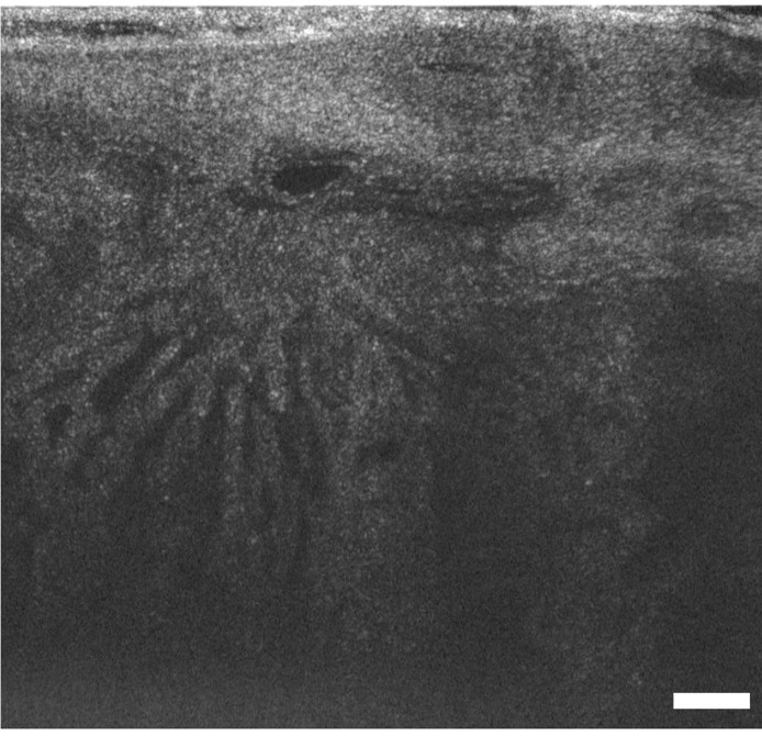
Non-invasive imaging allows us to examine skin structure and function without relying only on biopsy. We develop and apply optical imaging methods, including optical coherence tomography and KLEAR optical clearing, to visualize disease at cellular and tissue scales.
Selected projects
- Using OCT to detect and monitor skin cancer
- Increasing optical imaging depth with KLEAR
- Comparing non-invasive imaging with histology and 3D tissue pathology
Digital Signals
Skin as a Digital Sensor
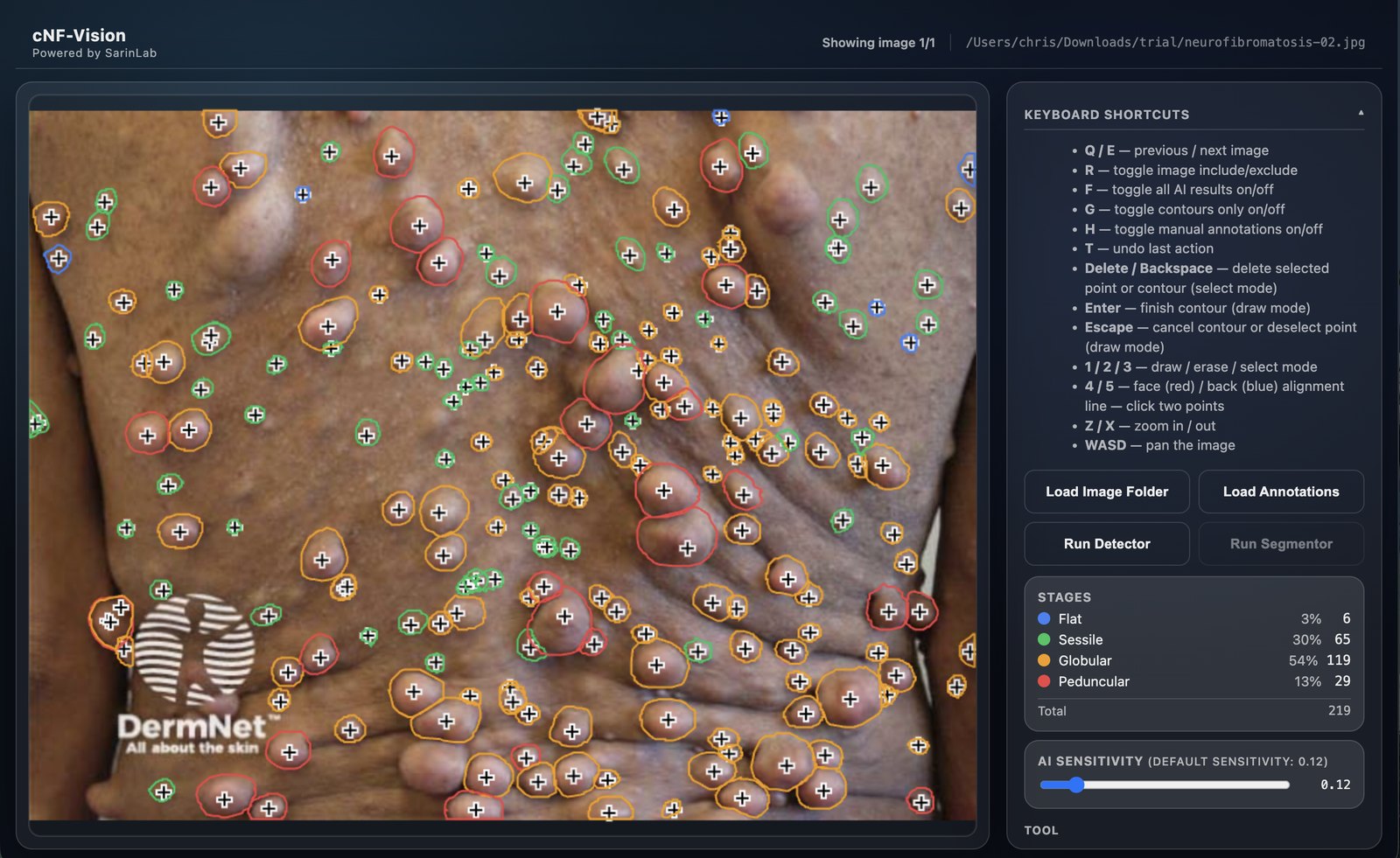
Digital tools can turn photographs and imaging data into quantitative measures of disease. We use artificial intelligence and image analysis to count lesions, measure disease burden, and develop more reliable outcomes for clinical research.
Selected projects
- Developing cNF-Vision to measure cutaneous neurofibroma burden
- Using longitudinal and whole-body imaging to track disease over time
- Automating the measurement of skin lesions and treatment response