
Technologies
We develop and apply technologies that allow us to see, measure, and interpret signals in the skin. Our work spans big data analysis, optical imaging, optical clearing, three-dimensional tissue analysis, spatial biology, and artificial intelligence.
Optical Imaging
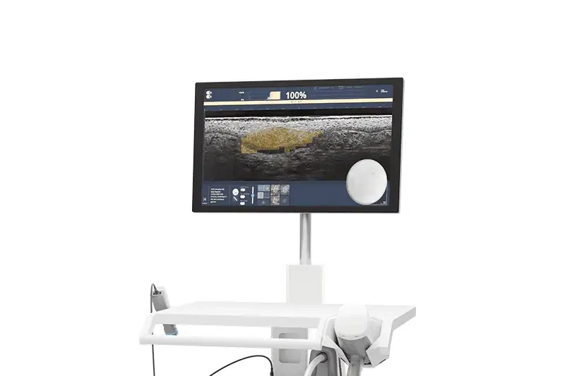
We use high-resolution optical imaging, including optical coherence tomography, to examine living skin without removing tissue.
What it reveals: Skin structure and light scattering at near-cellular resolution.
Applications
In collaboration with Yonatan Winetraub
Optical Clearing
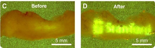
Optical clearing technologies use refractive index manipulation to reduce light scattering in skin, allowing visualization of deeper tissue structures.
What it reveals: Structures below the usual imaging depth of standard optical methods.
Applications
In collaboration with Guosong Hong
3D Molecular Pathology
We combine tissue staining, three-dimensional imaging, and computational reconstruction to study intact skin and tumor architecture.
What it reveals: The spatial organization of cells, structures, and molecular signals within tissue.
Applications
In collaboration with Jonathan Liu
Spatial Biology & Molecular Discovery
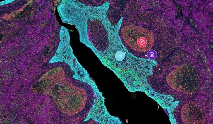
We use spatial transcriptomics, single-cell analysis, multiplex imaging, and molecular profiling to study how cells and biomarkers are organized within skin tissue.
What it reveals: The location and relationships of molecular and cellular signals within tissue.
Applications
AI & Computational Analysis
We use artificial intelligence and quantitative image analysis to measure disease from clinical photographs, optical images, and pathology data.
What it reveals: Reproducible measurements of lesion number, disease burden, and treatment response.
Applications
In collaboration with Olivier Gevaert